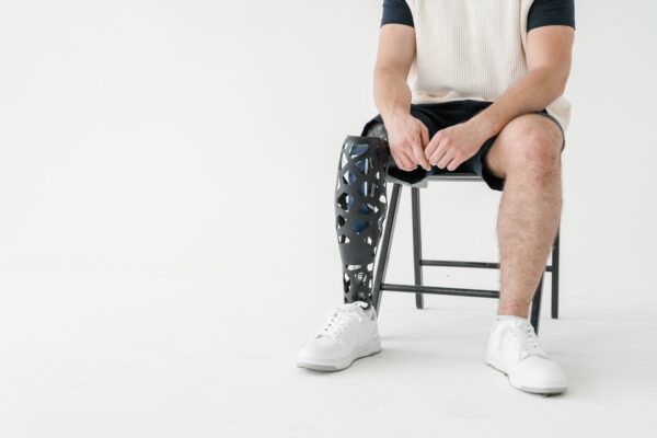
Ask any experienced prosthetist what the most common reason is for a patient abandoning their prosthesis, and you will get the same answer: socket discomfort. A prosthetic limb that causes pain, skin breakdown, or even mild chronic irritation will eventually be left in the corner of the bedroom, no matter how sophisticated its knee or foot component. Getting the socket right is not just a technical step in the fitting process — it is the single most important determinant of whether a patient actually uses their prosthesis.
This article looks at the principles of pressure management in lower-limb prosthetic sockets, with a focus on the practical realities of clinical practice in Pakistan.
Why Socket Fit Is So Challenging
The residual limb is a dynamic structure. It changes volume throughout the day — typically shrinking by the afternoon as fluid redistributes with gravity and activity. It changes over weeks and months as post-amputation oedema resolves, soft tissue atrophies, and the patient’s weight fluctuates. It changes with temperature, activity level, and hydration. A socket that fits perfectly at eight in the morning may be loose and uncomfortable by three in the afternoon.
At the same time, the residual limb is not engineered to bear weight. The tissues that envelop it — skin, subcutaneous fat, muscle, bone — have different mechanical properties and different tolerances for pressure. Bone prominences such as the fibular head, tibial crest, tibial condyles, and distal femur are pressure-intolerant and must be relieved. Other areas, such as the patellar tendon, the medial tibial flare, and the gluteal musculature, can tolerate and even benefit from controlled loading.
The prosthetist’s job is to design a socket that distributes load across pressure-tolerant areas while carefully relieving pressure-intolerant ones — and to do this for a residual limb that will not sit still.
Key Pressure-Tolerant and Pressure-Intolerant Areas
Transtibial (Below-Knee) Socket
Pressure-tolerant:
- Patellar tendon (the classic PTB loading zone)
- Medial tibial flare
- Popliteal fossa (with soft tissue present)
- Anterior-lateral surface (tibia + fibula, avoiding the tibial crest and fibular head)
Pressure-intolerant:
- Tibial crest and tibial tubercle
- Fibular head (common peroneal nerve)
- Distal tibia and fibula (bony end-bearing, unless the limb is specifically prepared for it)
- Hamstring tendons medially and laterally
Transfemoral (Above-Knee) Socket
Pressure-tolerant:
- Ischial containment area (ischial tuberosity and ramus)
- Gluteus maximus
- Lateral femoral shaft
- Anterolateral thigh (rectus femoris, vastus lateralis)
Pressure-intolerant:
- Distal femur (unless truly end-bearing)
- Greater trochanter
- Perineum (common site of excessive pressure in poor-fitting sockets)
- Adductor longus tendon (medial)
Knowing these zones is the starting point. Applying that knowledge to a real residual limb, under real loading conditions, is where clinical skill develops.
The Fitting Process: What to Look For
Static Assessment
Begin with the patient seated. Don the liner (if used), then the socket. Ask the patient to weight-bear gently. Before anything else, look at the socket brim: is it level? Is there excessive pistoning when the patient shifts their weight? Is the skin around the brim blanching or gathering?
Check the distal fit. In a PTB socket, there should be no more than one to two centimetres of space at the distal end, and ideally the residual limb should make gentle contact with the distal pad. A large distal gap creates a suction effect that draws the soft tissues distally, causing oedema, skin changes, and eventually breakdown.
Dynamic Assessment
Watch the patient walk. Look for these tell-tale signs of poor fit:
Pistoning: The socket rises during swing phase and descends in stance. This creates shear forces on the residual limb and indicates the socket is too large or the suspension system is inadequate.
Skin changes at the brim: Redness, chafing, or skin folds at the proximal brim usually indicate the brim is too high, too sharp, or incorrectly trimlined.
Mediolateral instability: The knee deviates medially or laterally during stance. Often caused by a socket that is too wide, or one that lacks adequate mediolateral bracing.
Limb positioning in the socket: The residual limb should sit consistently in the socket. If the patient can be seen hiking their pelvis or shifting their trunk to compensate for a loose socket, the fit needs attention.
Pressure Mapping
Where pressure mapping technology is available — and it is becoming increasingly affordable — it can be invaluable for confirming clinical impressions. Pressure-sensitive insoles or pads placed inside the socket provide real-time data on where load is distributed and can reveal pressure concentrations that are not apparent on visual inspection alone.
Managing Pressure Problems
Hot Spots and Skin Breakdown
A hot spot — a localised area of redness that persists for more than twenty minutes after removing the socket — is a warning. Skin breakdown follows. The first step is to identify whether the problem is in the socket itself (a pressure concentration due to shape) or related to the liner, suspension, or volume management.
Spot-grinding the socket at the offending area is the most direct solution for a rigid socket. In thermoplastic sockets, heat can be used to locally push out the problem area. For minor issues, strategic padding in adjacent areas can redistribute load away from the hot spot.
Volume Management
Volume fluctuation is best managed through sock management in patients without liners. A simple but systematic approach — adding or removing socks of known ply throughout the day based on the feel of the socket — allows the patient to maintain a consistent fit as volume changes.
For patients using pin-lock or elevated vacuum suspension, volume fluctuation is partially managed by the suspension system itself. Elevated vacuum systems are particularly effective at managing volume changes and reducing pistoning, though they add cost and complexity.
Residual limb wrapping with elastic bandage or a shrinker sock at the end of the day reduces oedema and slows volume loss — an important habit to establish early in the rehabilitation process.
Educating Your Patient
The patient is your partner in pressure management. They need to understand what normal discomfort looks like (the mild pressure of a new socket adjusting) versus what requires urgent attention (sharp pain, numbness, or skin that does not recover its normal colour within twenty minutes). Clear, practical education — ideally with written instructions and a demonstration — makes an enormous difference to outcomes.
In a busy outpatient clinic, it is tempting to rush through this. But the ten minutes you spend teaching a patient how to manage their own skin and volume will prevent many more clinic visits later.
Common Mistakes and How to Avoid Them
Casting in non-weight-bearing: Casting or scanning the residual limb in a non-weight-bearing position captures the limb in a relaxed state that does not reflect its shape under load. Where possible, cast or scan in partial or full weight-bearing. If non-weight-bearing casting is unavoidable, anticipate the changes that occur under load and modify accordingly.
Ignoring the distal end: Many prosthetists focus correctly on the brim and suspension but pay insufficient attention to the distal fit. A socket that ends one centimetre short of the residual limb creates a suction pocket that will slowly damage the distal tissue.
Over-padding hot spots directly: Padding directly on a hot spot increases pressure at that spot. Padding adjacent to a hot spot — a donut configuration — reduces pressure at the problem area.
Not reviewing fit after volume loss: Volume loss in the first three to six months post-amputation can be substantial. A socket that fitted well at six weeks may be significantly oversized by six months. Regular follow-up and early socket replacement or adjustment is essential, not optional.
A Word on Resources
OPPAK Academy recognises that many clinicians in Pakistan work with limited resources. Many of the assessment techniques described here — visual inspection, patient interview, systematic gait observation — require no special equipment. They require attention, a systematic approach, and the habit of looking for problems before they become crises.
Our clinical skills workshops and online modules include hands-on socket modification training, and our resource library includes fabrication guides that are designed for the equipment and materials available in Pakistan.
This article is part of the OPPAK Academy Blog series on clinical skills for orthotists and prosthetists. For questions or to share your experience managing socket fit in your practice, contact us at Contact@oppak.org.
Back to Blog